This content has been reviewed for medical accuracy. Always consult a qualified healthcare professional before making any medical decisions. [Last reviewed: 2026-02-10]
A comprehensive guide to understanding, detecting, and treating skin cancer
Skin cancer is the most common form of cancer worldwide, affecting millions of people each year. While the diagnosis can be concerning, early detection and modern treatment options have made skin cancer one of the most treatable forms of cancer. Understanding the different types, recognizing warning signs, and knowing when to seek medical attention can significantly improve outcomes and save lives.
This comprehensive guide will help you understand everything you need to know about skin cancer, from risk factors and prevention to the latest treatment advances.
What is Skin Cancer?
Skin cancer occurs when skin cells undergo abnormal growth, typically due to DNA damage caused by ultraviolet (UV) radiation from the sun or tanning beds. The skin is the body’s largest organ and consists of multiple layers. Cancer can develop in any of these layers, with different types arising from different cell types.
The three main layers of skin are:
Epidermis: The outermost layer, containing melanocytes (pigment-producing cells), squamous cells, and basal cells
Dermis: The middle layer, containing blood vessels, nerves, and hair follicles
Hypodermis: The deepest layer, primarily composed of fat and connective tissue
Most skin cancers develop in the epidermis, where cells are most exposed to UV radiation.
Types of Skin Cancer
There are three main types of skin cancer, each with distinct characteristics, behaviors, and treatment approaches:
1. Basal Cell Carcinoma (BCC)
Most Common: Basal cell carcinoma accounts for approximately 80% of all skin cancer cases, making it the most frequently diagnosed type.
Key Characteristics:
Develops in the basal cells of the epidermis
Grows slowly and rarely spreads (metastasizes) to other parts of the body
Most often appears on sun-exposed areas: face, ears, neck, scalp, shoulders, and back
Highly treatable with cure rates exceeding 95% when detected early
Can be locally destructive if left untreated
Appearance: Basal cell carcinoma may appear as:
A pearly or waxy bump with visible blood vessels
A flat, flesh-colored, or brown scar-like lesion
A bleeding or scabbing sore that heals and returns
A pink growth with a slightly elevated rolled border and crusted indentation in the center
2. Squamous Cell Carcinoma (SCC)
Second Most Common: Squamous cell carcinoma accounts for approximately 20% of skin cancer cases and is more aggressive than basal cell carcinoma.
Key Characteristics:
Arises from squamous cells in the outer layer of skin
Grows faster than basal cell carcinoma
Can metastasize to lymph nodes and other organs if left untreated
Commonly appears on sun-exposed areas but can develop anywhere
May arise from actinic keratoses (pre-cancerous lesions)
Cure rate of 95% when caught early; drops significantly if it spreads
Appearance: Squamous cell carcinoma may present as:
A firm, red nodule or bump
A flat lesion with a scaly, crusted surface
A rough, scaly patch that may bleed
A wart-like growth
An open sore that doesn’t heal
A raised growth with a central depression
3. Melanoma
Most Dangerous: Although melanoma accounts for only about 1% of skin cancer cases, it causes the vast majority of skin cancer deaths due to its ability to spread rapidly.
⚠️ Critical Information:
Melanoma develops in melanocytes (pigment-producing cells)
Can spread quickly to lymph nodes and internal organs
When detected early (Stage 0 or I), the 5-year survival rate exceeds 99%
If it spreads to distant organs, survival rates drop significantly
Can develop in existing moles or appear as new pigmented or unusual-looking growths
More common in people with fair skin, but can affect all skin types
Appearance: Melanomas can vary widely but often show the ABCDE warning signs (detailed below):
A dark brown or black spot or mole that changes
A mole with irregular borders, multiple colors, or asymmetry
A new pigmented or unusual-looking growth
A spot that itches, bleeds, or feels different
Less Common Types
Other, rarer forms of skin cancer include:
Merkel Cell Carcinoma: Rare, aggressive cancer appearing as firm, painless nodules on sun-exposed skin
Kaposi Sarcoma: Develops in skin blood vessels, common in immunocompromised individuals
Sebaceous Gland Carcinoma: Aggressive cancer originating in oil glands, often on the eyelids
Dermatofibrosarcoma Protuberans: A rare cancer growing in the dermis layer
Risk Factors for Skin Cancer
Understanding risk factors can help you take preventive measures and remain vigilant about skin changes:
Controllable Risk Factors:
UV Exposure: Excessive sun exposure, especially sunburns during childhood, significantly increases risk
Tanning Beds: Indoor tanning dramatically increases melanoma risk, particularly when started before age 30
Lack of Sun Protection: Not using sunscreen, protective clothing, or shade
Occupational Exposure: Jobs requiring prolonged outdoor work without protection
Non-Controllable Risk Factors:
Fair Skin: Light skin, hair, and eye color (though skin cancer affects all skin types)
Family History: Having close relatives with skin cancer or melanoma
Personal History: Previous skin cancer diagnosis increases risk of recurrence
Many Moles: Having more than 50 moles or atypical (dysplastic) moles
Age: Risk increases with age, though melanoma is increasingly common in younger adults
Weakened Immune System: Due to disease or medications
Certain Genetic Conditions: Such as xeroderma pigmentosum or familial atypical multiple mole melanoma (FAMMM) syndrome
Signs and Symptoms: The ABCDE Rule
Early detection is crucial for successful treatment. The ABCDE rule is a simple guide to identifying potentially cancerous moles and spots:
Letter
Stands For
What to Look For
A
Asymmetry
One half of the mole doesn’t match the other half. Normal moles are typically symmetrical.
B
Border
Irregular, scalloped, or poorly defined edges. Healthy moles have smooth, even borders.
C
Color
Varied colors within the same mole (brown, black, tan, red, white, or blue). Normal moles are typically one shade.
D
Diameter
Larger than 6mm (about the size of a pencil eraser), though melanomas can be smaller.
E
Evolving
Changes in size, shape, color, elevation, or symptoms (itching, bleeding, crusting).
⚠️ See a Doctor Immediately If You Notice:
A new spot, mole, or growth that looks different from others
A sore that doesn’t heal within 3-4 weeks
A spot that continues to itch, hurt, crust, scab, or bleed
Any mole or spot that exhibits the ABCDE warning signs
A mole that looks significantly different from your other moles (“ugly duckling sign”)
Diagnosis of Skin Cancer
If your doctor suspects skin cancer, they will follow a systematic diagnostic approach:
1. Visual Examination
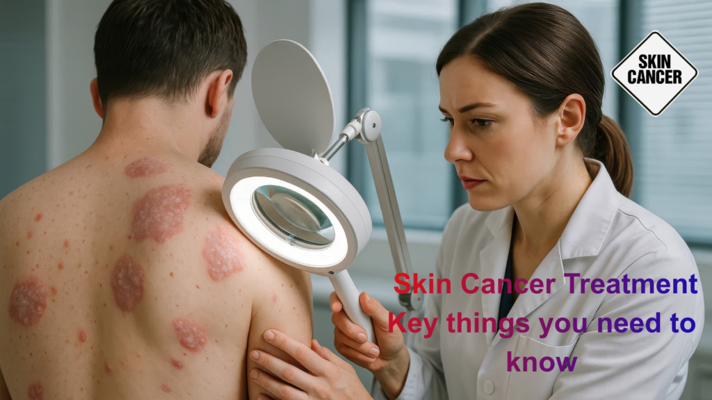
Your doctor will examine the suspicious area and check your entire body for other concerning spots. They may use:
Dermoscopy: A handheld device with magnification and light to examine skin lesions more closely
Full Body Skin Exam: Checking all areas of skin, including scalp, between toes, and other hidden areas
2. Biopsy
If a lesion appears suspicious, a biopsy is performed to confirm the diagnosis. Types include:
Shave Biopsy: The top layers of skin are shaved off with a small blade
Punch Biopsy: A circular tool removes a deeper sample of skin
Excisional Biopsy: The entire lesion plus a margin of normal skin is removed
Incisional Biopsy: Part of the lesion is removed for examination
The tissue sample is sent to a pathology lab where specialists examine it under a microscope to determine if cancer cells are present and, if so, what type.
3. Staging (for Melanoma and Advanced Cases)
If cancer is confirmed, additional tests may be needed to determine the stage:
Sentinel Lymph Node Biopsy: Determines if cancer has spread to nearby lymph nodes
Imaging Studies: CT scans, MRI, or PET scans to check for spread to other organs
Blood Tests: Including LDH levels, which may be elevated in advanced melanoma
Melanoma Staging:
Stage 0 (in situ): Cancer cells only in the top layer of skin
Stage I: Thin melanoma has not spread
Stage II: Thicker melanoma, has not spread
Stage III: Spread to nearby lymph nodes
Stage IV: Spread to distant organs or skin areas
Treatment Options
Treatment for skin cancer depends on the type, size, location, stage, and overall health. Modern medicine offers multiple effective approaches:
Surgical Treatments
1. Excisional Surgery
The most common treatment for skin cancer is. The cancerous tissue and a margin of healthy tissue are removed. For basal and squamous cell carcinomas, this is often curative.
2. Mohs Micrographic Surgery
A specialized technique where thin layers of cancer-containing skin are progressively removed and examined until only cancer-free tissue remains.
Benefits:
Highest cure rate (up to 99% for first-time BCC)
Minimizes the removal of healthy tissue
Ideal for cancers on the face, ears, and other cosmetically sensitive areas
Excellent for recurrent cancers or those with poorly defined borders
3. Curettage and Electrodesiccation (C&E)
The cancer is scraped away with a curette, then heat is applied to destroy remaining cancer cells and control bleeding. Best for small, low-risk basal and squamous cell carcinomas.
4. Cryosurgery
Liquid nitrogen is used to freeze and destroy abnormal cells. Effective for precancerous lesions and some small, superficial skin cancers.
Non-Surgical Treatments
1. Radiation Therapy
High-energy beams target and kill cancer cells. Used when:
Surgery is not an option
Cancer is in a difficult location
Additional treatment is needed after surgery
Patient cannot undergo surgery due to health conditions
2. Topical Medications
Prescription creams or gels applied directly to the skin:
5-Fluorouracil (5-FU): Chemotherapy cream for superficial basal cell carcinomas and precancerous lesions
Imiquimod: Immune response modifier that stimulates the immune system to fight cancer cells
3. Photodynamic Therapy (PDT)
A light-sensitizing medication is applied to the skin, then activated with a special light to destroy cancer cells. Effective for actinic keratoses and some superficial skin cancers.
Advanced Treatments for Melanoma
1. Immunotherapy
Breakthrough treatments that harness the immune system to fight cancer:
Checkpoint Inhibitors: Drugs like pembrolizumab (Keytruda), nivolumab (Opdivo), and ipilimumab (Yervoy) that help the immune system recognize and attack cancer cells
Effectiveness: Significantly improved survival rates for advanced melanoma
Used for: Stage III and IV melanoma, and as adjuvant therapy after surgery
2. Targeted Therapy
For melanomas with specific genetic mutations (BRAF, MEK, KIT):
Can rapidly shrink tumors, but resistance may develop
3. Chemotherapy
Traditional chemotherapy may be used for advanced melanoma when other treatments haven’t worked, though it’s less common now due to more effective immunotherapy and targeted therapy options.
4. Tumor-Infiltrating Lymphocyte (TIL) Therapy
An emerging treatment in which immune cells are extracted from the tumor, expanded in vitro, and infused back into the patient to fight cancer.
Combination and Sequential Therapies
Many patients receive multiple treatments, either simultaneously or sequentially, to achieve optimal results. Your oncologist will create a personalized treatment plan based on your specific situation.
Prevention: Protecting Your Skin
While not all skin cancers can be prevented, you can significantly reduce your risk with these evidence-based strategies:
Sun Protection Essentials
Daily Sun Protection (365 Days a Year):
Sunscreen: Use broad-spectrum SPF 30+ daily, even on cloudy days
Apply 1 ounce (enough to fill a shot glass) to cover your body
Reapply every 2 hours, or immediately after swimming or sweating
Don’t forget ears, neck, hands, and the tops of feet
Seek Shade: Especially between 10 AM and 4 PM when UV rays are strongest
Wear Protective Clothing:
Long-sleeved shirts and long pants
Wide-brimmed hat (3-inch brim all around)
UV-blocking sunglasses
Consider UPF-rated clothing for extra protection
Additional Prevention Strategies
Avoid Tanning Beds: Indoor tanning increases melanoma risk by 75% when started before age 30
Check Your Skin Monthly: Perform self-examinations to catch changes early
Annual Dermatology Visits: Especially if you have risk factors
Protect Children: One blistering sunburn in childhood doubles melanoma risk
Know Your Medications: Some drugs increase sun sensitivity
Examine Your Scalp: Use a blow dryer and a mirror or ask someone to help
Living with and After Skin Cancer
Follow-Up Care
After treatment, regular follow-up is essential:
Skin Checks: Every 3-12 months, depending on cancer type and stage
Self-Examinations: Monthly checks of your entire skin surface
Imaging: For melanoma patients, periodic scans to monitor for recurrence
Lymph Node Checks: Physical examination and sometimes an ultrasound
Emotional Support
A cancer diagnosis affects mental and emotional wellbeing. Consider:
Counseling or therapy
Support groups for skin cancer survivors
Open communication with loved ones
Stress management techniques
Reducing Recurrence Risk
Strict sun protection for life
Regular dermatologist visits
Healthy lifestyle: diet, exercise, no smoking
Vitamin D supplementation if avoiding sun (consult your doctor)
When to See a Doctor
Seek Medical Attention If:
You notice any new, changing, or unusual skin growths
A sore doesn’t heal within 3-4 weeks
You have a mole that exhibits any ABCDE warning signs
You have significant risk factors and haven’t had a skin check
You’ve had skin cancer before
You notice swollen lymph nodes
You experience unexplained symptoms like persistent cough, bone pain, or headaches (if you’ve had melanoma)
The Future of Skin Cancer Treatment
Research continues to advance our understanding and treatment of skin cancer:
AI-Assisted Diagnosis: Artificial intelligence helping dermatologists detect cancer earlier and more accurately
Liquid Biopsies: Blood tests to detect circulating tumor DNA for early detection and monitoring
Vaccine Development: Therapeutic vaccines being studied for melanoma prevention and treatment
Combination Immunotherapies: Novel combinations showing promise for resistant cancers
Precision Medicine: Tailoring treatment based on individual tumor genetics
Improved Targeted Therapies: New drugs targeting different genetic mutations
Conclusion
Skin cancer is highly treatable when detected early, with cure rates exceeding 95% for basal and squamous cell carcinomas and 99% for early-stage melanoma. The key to successful outcomes lies in prevention, early detection, and prompt treatment.
By protecting your skin from UV radiation, performing regular self-examinations, and maintaining annual dermatology appointments, you can significantly reduce your risk and catch any problems early. If you notice any suspicious changes in your skin, don’t wait—see a dermatologist promptly.
Remember, when it comes to skin cancer, early detection saves lives. Your skin tells a story—make sure you’re paying attention to what it’s saying.
Medical Disclaimer:
This article is for informational purposes only and should not be considered medical advice. Skin cancer diagnosis and treatment should always be conducted by qualified healthcare professionals. If you have concerns about skin changes or risk factors, please consult with a board-certified dermatologist or oncologist. Treatment decisions should be made in consultation with your medical team based on your specific circumstances, medical history, and current health status.
Need Expert Medical Care?
Medsmonk connects patients with world-class dermatologists and oncologists for skin cancer screening, diagnosis, and treatment. Our medical tourism services provide access to cutting-edge treatments and personalized care.
Contact Medsmonk today to learn more about your options.
Resources
Resources
World Health Organization (WHO) – Ultraviolet radiation and health (UV risk & prevention): Click here
American Cancer Society – Basal & squamous cell skin cancers (overview): Click here
National Cancer Institute (NCI) – Melanoma Treatment (PDQ®): Click here
American Academy of Dermatology (AAD) – ABCDEs of melanoma (warning signs): Click here
Centers for Disease Control and Prevention (CDC) – Ultraviolet (UV) radiation facts: Click here
Medical Review DisclosureThis content has been reviewed for medical accuracy. Always consult a qualified healthcare professional before making any medical
Need this medicine?
Our pharmacists can confirm availability, pricing and what your country requires for import.